Diagnostic Imaging
Atlanta Imaging Center
Trying to diagnose a disease, injury, or sickness without diagnostic imaging is like searching for buried treasure without a treasure map. It’s like taking a shot in the dark, completely blind, and simply hoping for the best results. Here at AICA Orthopedics, in our Atlanta imaging center, we don’t want to guess with your health, so we utilize advanced diagnostic imaging technology and diagnostic radiology to diagnose, treat, and evaluate the progress of our patients.
If you’re having chronic headaches or debilitating knee pain, we could definitely start a list of possibilities without exploring the discomfort further. But we want to know exactly what the issue is, where it’s coming from, and why it’s happening. That way, we can treat it accordingly and give you the best chance at recovery, pain-relief, and optimal health and wellness. Our diagnostic imaging services are just one way that we provide the best comprehensive care for our patients.
Advanced Dianostic Imaging Equipment
Here at AICA Orthopedics, we have only the best diagnostic imaging equipment. It is the most advanced, most up-to-date, and most effective equipment available today in medical imaging. At our Atlanta imaging center we can run one test or several, depending on what your situation necessitates. We will explore all possibilities for the cause of injury or disease before we begin our imaging diagnostics so that we make the most of our time and yours, and so that we can fully explore every possibility for the root cause of your discomfort. While we have other diagnostic procedures available should we need them, our imaging equipment helps us diagnose a large number of injuries and ailments so that we can properly treat you.
MRI
CT Scan
X-Ray
Fluoroscopy
Experienced Imaging Specialists
Our diagnostic imaging specialists are highly qualified, extremely educated, and very experienced. They make your diagnosis, care, treatment, and recovery their top priority, and they will always listen to any concerns or fears that you have regarding an imaging procedure. Each specialist utilizes imaging tools to best meet their patients’ needs and pinpoint the exact cause of pain or injury.
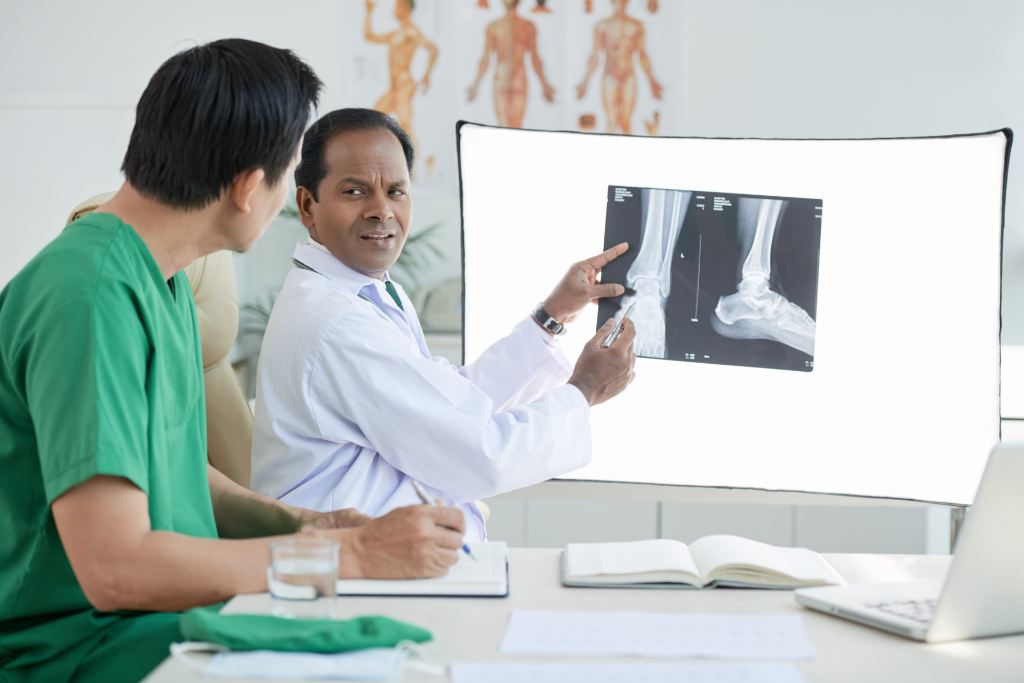
Orthopedic specialists use various diagnostic imaging tests for diagnosis. X-rays are the most common form of imaging but also the most basic. An X-ray can be used to diagnose bone fractures, arthritis, or joint issues such as dislocation or space. Different X-rays at certain positions can show the problem area from various angles.
A CT scan or a Computed Tomography scan is much like an X-ray; however, it provides a three-dimensional view to the body and provides great angles to bones and joints, unlike an x-ray. The downside to a CT scan is that they can be more time consuming than that of an x-ray, and they also give off more radiation. Although a CT scan is three dimensional, it does not provide a clear picture of ligaments and tendons, which can make diagnosing soft tissue injuries difficult.
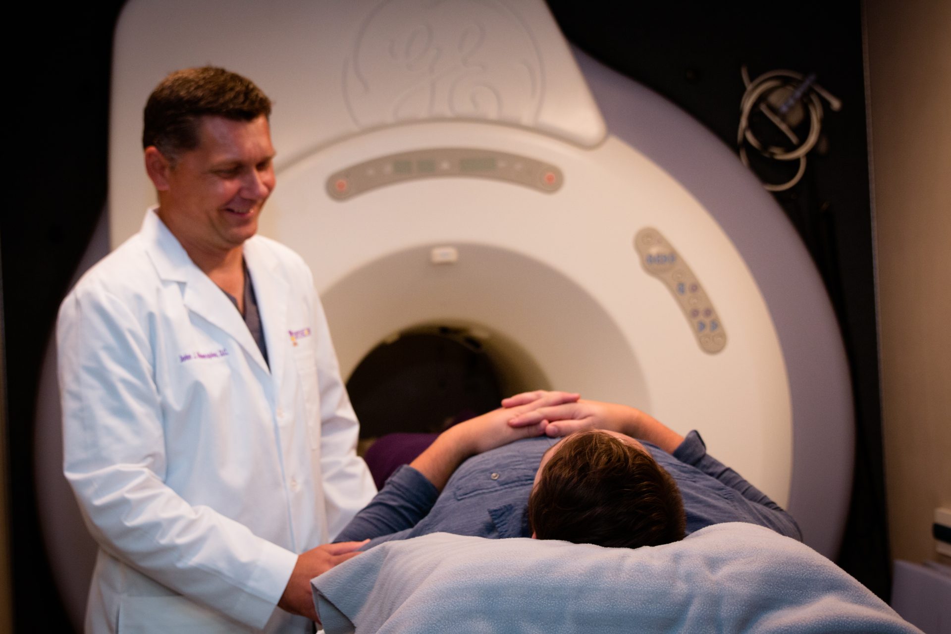
Magnetic Resonance Imaging (MRI) is often used to evaluate soft tissue. With this test, the patient will lie down in a machine surrounded by a magnetic field. The test creates three-dimensional pictures when the magnetic field causes the molecules in the body to vibrate. The plus side to this test is that it provides detailed images of the body as well as no exposure to radiation. Some patients, however, have concerns with this form of diagnostic testing due to fears of claustrophobia. Still, our orthopedic doctors will consider your interests and make sure that your experience is one that leaves you feeling comfortable and relaxed.
An EMG test is used to examine the electrical activity of muscles and assess the function of nerves. The activity between the resting and active state is measured to look for problem areas.
A neurologist focuses on the nerves and central nervous system. To assess these types of issues, neurologists also use imaging, sometimes differently than an orthopedic specialist. For neurological issues, EMG imaging can stimulate muscle nerves to detect abnormalities. ENG testing is used to evaluate vertigo and vision issues. MRI scans use a large magnet and radio frequencies to produce images of the brain and spine. X-rays of the skull, neck, or back can be used to diagnose tumors or bone injuries, arthritis, and disc degeneration.
For spinal pain, interventional spine specialists can use an MRI scan to get a clear picture of the spine and look for problem areas. When the MRI is inconclusive, a discogram is used specifically for looking at the health of each vertebra to determine if a specific disc is the cause of spinal pain. Fluoroscopy is an imaging technique used to insert dye into the vertebra that then projects X-ray images onto a monitor. The dye helps spine specialists search for leaks that could be areas of concern. This type of dye can also be used with a CT scan to reveal tears, bulges, or scarring in specific areas of the spine.
Chiropractors focus on the health of the central nervous system in conjunction with the musculoskeletal system. Chiropractors at AICA Orthopedics first perform a physical examination examining your spine or other parts of the body that are causing pain. If they can’t get a clear picture of the problem this way, then they can use X-rays and MRI imaging for a more specific picture of the areas that are the source of pain.
If you have an injury or are experiencing pain, our specialists at AICA Orthopedics are equipped to help. To learn more about our imaging services, click on the links in the menu bar or contact one of our many Atlanta offices today.


