In addition to the pain you may experience as a result of nerve damage, it can also be frustrating to try and diagnose the source of the issue. You may have undergone a number of tests and spoken to many specialists to try and address your pain. Because there are over one hundred types of nerve damage, it can be difficult to pin down the symptoms you are experiencing. An experienced orthopedic surgeon will be able to work with you to understand your condition- one of the ways they do this may be to schedule you for an MRI scan. But will this imaging show actual nerve damage, or even a pinched nerve? Read on to find out.
Damaged and Pinched Nerves
Though they are small and hard to identify, nerves power our entire body. There are three main types of nerves operating within us at all times. Autonomic nerves control our involuntary mechanisms like blood pressure, heart rate, and digestive processes. Motor nerves, on the other hand, control voluntary movement and actions by communicating to muscles from the brain and spinal cord. Sensory nerves then communicate from the skin and muscles back to your brain and spinal cord, causing you to feel pain. These nerves can all be damaged by sports injuries, falls, or accidents. Depending on which nerves have been impacted, your symptoms may differ.
One common cause of nerve pain is a pinched nerve, in which it is compressed between tissue such as bones, tendons, or ligaments. This pressure causes inflammation in the nerve, leading to pain and discomfort. Often, misalignment in the upper spine and neck causes this condition as a result of repetitive motion, poor posture or sleeping positions, stress, or simply aging.
Nerve damage and pinched nerves may be the cause if you are experiencing numbness and decreased sensation, sharp or burning pain, or a tingling sensation in your extremities. Weakness and problems with motor skills are also signs of an issue with nerve damage. These symptoms can all become worse over time without proper care from an orthopedic surgeon.
MRIs for Diagnosing Nerve Pain
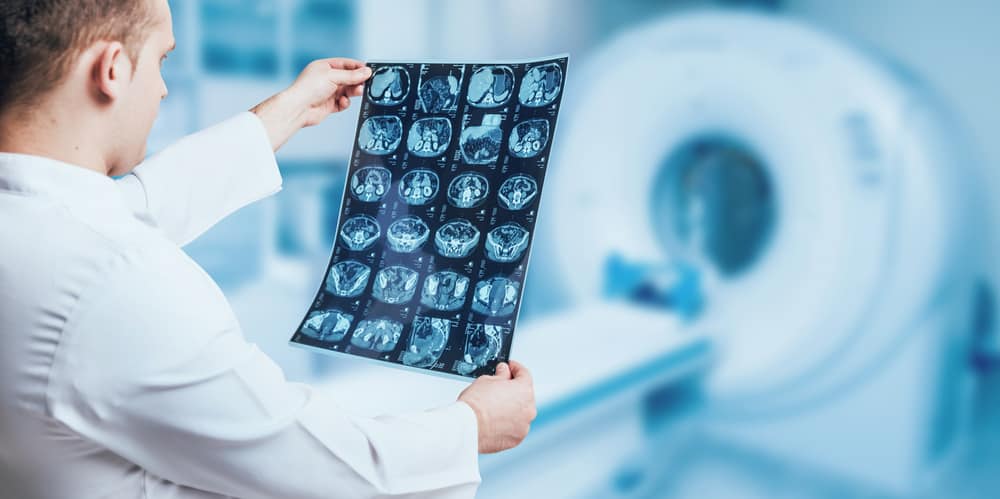
An MRI, or Magnetic Resonance Imaging, is a scan that is able to render images of soft tissue structures throughout the body. This allows physicians to view a patient’s full spinal anatomy in order to determine the cause of a patient’s pain, which can then be correlated to symptoms to provide a diagnosis.
MRIs are able to provide such in-depth prognostic information by using a combination of a strong magnetic field, radio waves, and a computer that is able to provide these extremely detailed pictures. The power of these factors means doctors can see not only the spine, but individual vertebrae, discs, the spinal cord, and even the small spaces between vertebrae where nerves pass. This image can be viewed on a computer or printed for evaluation, and it is known to be the most sensitive imaging test currently available for the spine.
What to Expect at Your MRI
If you are experiencing symptoms consistent with nerve damage, your orthopedic surgeon will likely recommend you sit for an MRI to further evaluate your condition. Some people are frightened by the prospect of getting an MRI because of the heavy machinery, but they actually come with fewer risks than other scans due to a lack of radiation exposure. You will also not need to use any medications to undergo an MRI and will be able to go about your day as normal immediately following the scan.
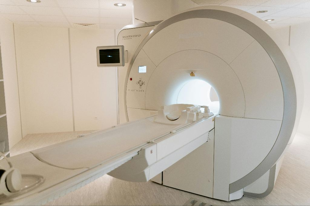
On the day of your MRI, you will be asked to remove any jewelry or external medical devices to avoid interference with the magnetic fields. If you use a pacemaker or other non-removable device, you will need to discuss that with your doctor ahead of time. You will then be placed on a table that slides into a tube, where you will lie still as pictures are taken. There may be a speaker that can be used for communication with medical staff during this time. For some, being in the tube for a long time can cause discomfort or claustrophobia, which can also be discussed in advance.
At AICA Orthopedics, our imaging center is a part of our multidisciplinary medical facility, allowing your scans to be evaluated by the same orthopedic surgeons you are working with. In addition, you will have access to skilled pain management specialists and physical therapists to execute the recovery plan you develop with your doctor. As specialists in car accident injuries, the team at AICA Orthopedics has seen a wide range of possible nerve injuries, and we aim to create a safe and effective recovery plan for all our patients. Call us today.


